Spina Bifida is a congenital defect in which the vertebral column does not enclose properly, exposing the spinal cord. The degree of spinal cord involvement, from exposure to herniation, defines the severity and classification of the condition. In Spina Bifida Occulta and Meningocele defects, the exposure of the spinal cord is minimal, and patients present little to no symptoms. Myelomeningocele (MMC) and Myeloschisis (MS) defects are the most common and most severe. In both instances the spinal cord and nerves can be exposed to amniotic fluid. MMC and MS can result in partial or complete paralysis below the level of the spinal opening, inability to walk, and/or bladder and bowel dysfunction. The severity of these symptoms is associated with the continuous exposure of the neural tissues in utero. For this reason, fetal surgery was sought as the most effective way of preventing irreversible damage to the patient¹.


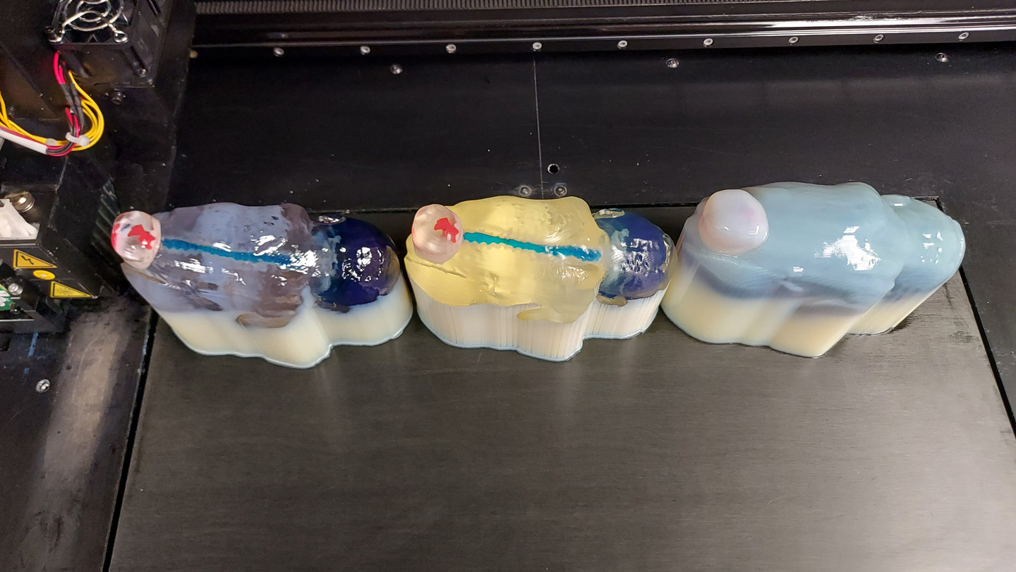
Considering the high-stakes nature of in-utero Spina Bifida repair, pediatric neurosurgeons at Orlando Health have partnered with DASH to produce patient-specific 3D printed models of the defect before surgery. Since early 2021, DASH has reconstructed and 3D printed twelve (12) spina bifida cases from MRI and 3D ultrasound.
Myelomeningocele
Myeloschisis
¹Copp, A., Adzick, N., Chitty, L. et al. Spina bifida. Nat Rev Dis Primers 1, 15007 (2015). https://doi.org/10.1038/nrdp.2015.7