Left Atrial Appendages (LAA) are small sacs in the muscle wall of the left atrium. While the presence of an LAA does not affect the proper function of the heart, studies have shown that most left atrium clots are formed in the LAA. The pouch-like structure of LAAs allows blood to pool and form clots. With each heartbeat, blood clots can travel from the LAA into peripheral vasculature and cause a stroke. Healthcare providers may recommend a LAA closure procedure to patients at risk of developing blood clots. In LAA closure procedures, cardiologists will seal off the appendage to prevent clots from forming and causing a stroke. The procedure will also eliminate the need for patients to take blood thinners to prevent the formation of clots altogether¹.
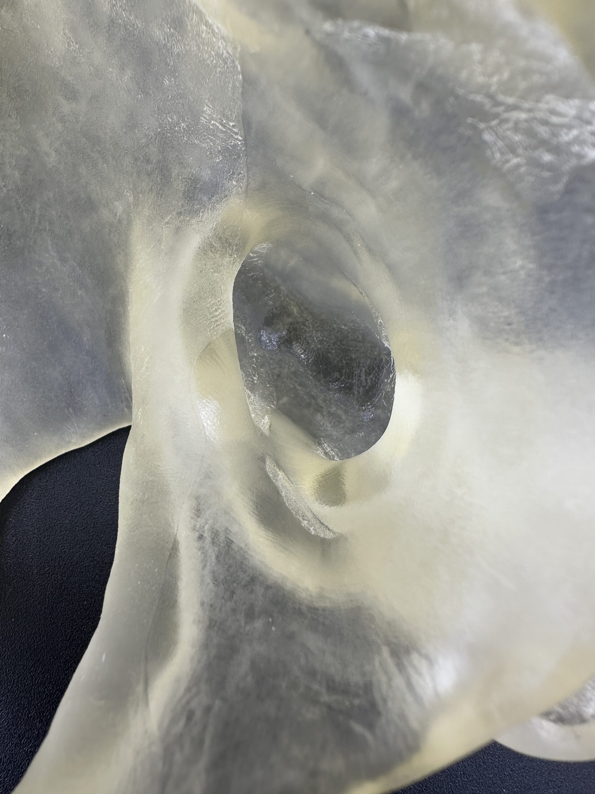

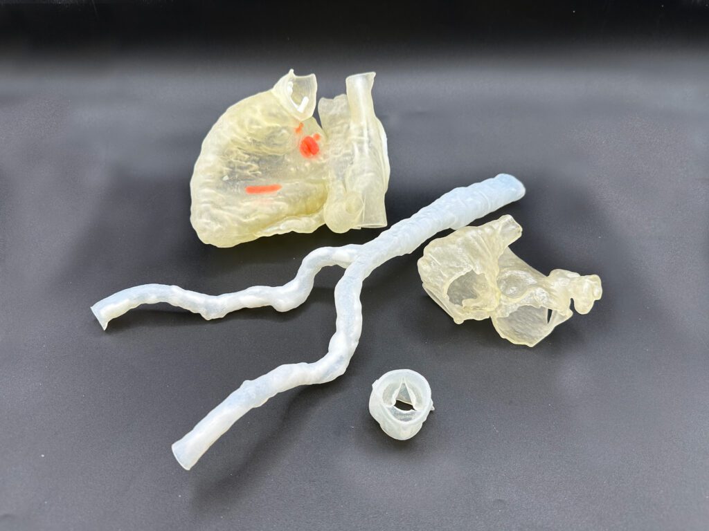
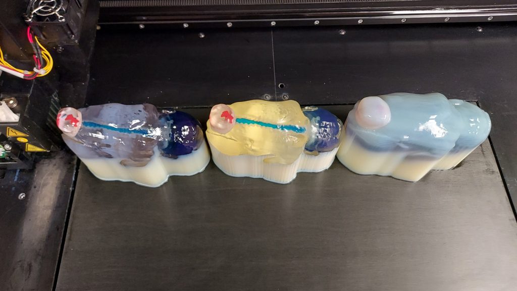
DASH is working with Orlando Health cardiologists and electrophysiologists to reconstruct and 3D print complex LAAs to plan and practice the procedure, in addition to testing the fit of closure devices. 3D models of the LAAs are 3D printed in flexible transparent materials for tactile feedback and visualization outside of the catheterization lab. LAA models are also printed on stands that approximate the correct anatomical position and orientation of the model.
LAA Case
¹Left Atrial Appendage Closure (LAA), Cleveland Clinic (https://my.clevelandclinic.org/health/treatments/17167-left-atrial-appendage–closure)